Correct!
Squamous Cell Carcinoma accounts for up to 95% of all cancers in the oral cavity. Therefore, it is most likely the case of of this patients oral lesion.
Results of Investigations:
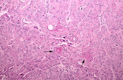
The biopsies show a well-differentiated squamous cell carcinoma of the tongue with lymph node metastasis:
|
| High power photomicrograph of a well differentiated squamous cell tumor with well-defined intercellular bridges and keratin pearls (black arrows). Nuclear pleomorphism is mild to moderate, and the mitotic rate is low. Courtesy of Kenneth Haines, MD. |
CT Head with Contrast shows a 3cm by 3.5 cm mass in the left side of the tongue involvement of deep tongue musculature. There is no extension of the tumour into the mandible and the tumour does not cross midline. The CT scan is also identified a 1.5 cm enlarged lymph node. No other masses were detected. This indicates a stage III cancer (T2, N1, M0).
| Clinical Staging of Oral Cancers |
Chest x-ray and liver tests were normal indicating that metastasis to lungs and liver are unlikely (M0).